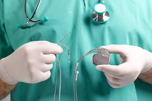
Insertion of a permanent pacemaker is a minimally invasive procedure. Access to heart chambers takes place as transvenous access to local anaesthesia. The most common method is via the subclavian vein or the cephalic vein. In rare cases, it is through femoral vein or the internal jugular vein. Either in an operating room or in a cardiac catheterization laboratory, the pacemaker implant procedure is performed.
In the infraclavicular region, the pacing generator is placed subcutaneously. Via thoracotomy, the pacemaker leads are implanted surgically. The pacing generator is then placed in the abdominal area. Either via left or right pectoral sites, single chamber and dual chamber insertion can be accomplished. The chest is then prepared. Sterile drapes are applied to the incision area to keep it as sterile as possible. Antibiotic prophylaxis is nowadays employed for the implantation. Preoperative antibiotic can reduce the chances of any infection by almost 80 percent. Cefazolin 1g is administered intravenously one hour prior to the procedure. Other antibiotics can be administered if the patient is allergic to cephalosporins, vancomycin, or penicillins.
The central vein is accessed percutaneously. Due to skeletal landmarks being deviated in some patients, there will be a need of fluoroscopic examination to reduce the time and complications in access. At the junction of first rib and the clavicle, the subclavian vein is typically accessed. For the confirmation of deep vein thrombosis, a phlebography is required for visualization of the vein.
Now a guide wire is advanced through the access needle and tip of the guide wire in placed in the right atrium or venacaval area under fluoroscopy. The guide wire is kept in place after the needle is withdrawn. If required, a second guide wire is also placed. Double wire technique may be employed through a sheath which is then withdrawn. Two separate sheaths can be manoeuvred over the two guide wires. During the lead advancement, some friction can be felt.
An incision of one to two inches is made in the area of the infraclavicle, which is parallel to the middle third of the clavicle and a subcutaneous pocket is made with both sharp and blunt dissection. This is for the implantation of the pacemaker generator. In many cases, surgeons prefer the access later and pocket first.
A peel-like special sheath and dilator are advanced over the guide wire. The guide wire and dilator are withdrawn keeping the sheath in place. A stylet is then inserted in the center channel of the pacemaker lead making it more rigid. This lead-stylet combination is then inserted into the sheath and advanced to the concerned heart chamber under fluoroscopy. In order to prevent dislodgement, the ventricular lead is positioned before the atrial lead. For the positioning in the tricuspid valve, a small curve at the tip of the stylet make it more mobile to reach the right ventricular apex. The introducing sheath is peeled once the lead is secured. With a pacing system analyser, the lead impedances are measured after the pacing lead stylet is removed. To prevent diaphragmatic stimulation, the pacing is performed at 10V.
After the confirmation of thresholds and lead position, the proximal end of the lead is secured to the pectoralis tissue with the help of a non-absorbable suture. This suture is sewn to a sleeve which is located on the lead. This is placed in the right atrium is a second lead is required. For patients who have already had a cardiac surgery, the lead tip is positioned medially or in the free lateral wall of right atrium. Same process of stylet withdrawal is followed after this. After positioning and testing of leads, the pacemaker pocket is fed with antimicrobial solution and the pulse generator is connected to the leads. To prevent migration or twiddler syndrome, many surgeons secure the generator to the underlying tissue with non-absorbable suture.
Before final confirmation of lead positioning, a look is taken under the fluoroscope. With the help of adhesive strips and absorbable sutures, the incision is closed. A sterile dressing is then applied on the surface. To limit movement for 12 to 24 hours, an immobilizer or arm restraint is applied. The chances of pneumothorax are ruled out with the help of a postoperative chest radiograph.


