Craniotomy (Brain Tumor)

Starts from $6700

Craniotomy

About Craniotomy (Brain Tumor)

Craniotomy (Brain Tumor) in India starts from $6700. The total cost of the treatment depends on the diagnosis and facilities opted by the patient.

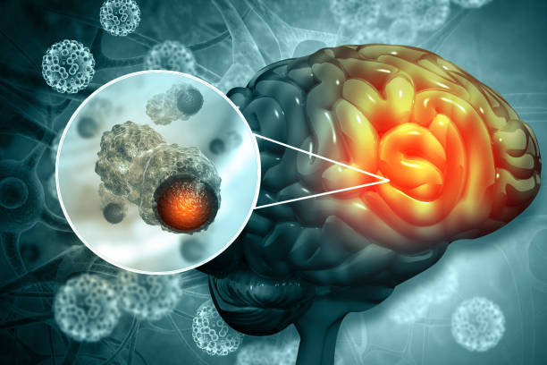
Craniotomy surgery is one of the most common types of brain surgery conducted to treat a brain tumor. It mainly aims at removing a lesion, tumor, or a blood clot in the brain by opening a flap above the brain to access the targeted area. This flap is removed on a temporary basis and again put in place when the surgery is done. Around 90 percent of the cases of brain tumors are diagnosed in adults aged between 55 and 65. Among children, a brain tumor is diagnosed within an age range of 3 to 12 years.

Craniotomy procedures are conducted with the help of magnetic resonance imaging (MRI) scans to reach the location precisely in the brain that requires treatment. A three-dimensional image for the same is achieved of the brain in conjunction with localizing frames and computers to view a tumor properly. A clear distinction is made between abnormal or tumor tissue and normal healthy tissue and to access the exact location of the abnormal tissue.

Frequently asked questions about Craniotomy (Brain Tumor)

  • An intravenous line is first placed in the arm and general anaesthesia is employed while the patient lies on the operating table.
  • When the patient is no more awake, the head is placed in a 3-pin skull fixing the device. This is attached to a table and keeps the head in a fixed position all throughout the procedure.
  • An insertion of a lumbar drain is made in the lower back to drain out the cerebrospinal fluid. A relaxation drug for the brain named mannitol can be administered at this stage.
  • With an antiseptic, the skull is prepared and an incision is made usually behind the hairline. A good cosmetic result is achieved after the surgery and a hair sparing technique, which requires shaving only one-fourth inch-wide area along the incision proposed is aimed. At times, the total area of the incision may be shaved.
  • The skin and muscles are then removed from the bone and folded back, burr holes are made in the skull using a drill.
  • A saw is introduced through the holes made and cuts the bone flap’s outline. The protective covering of the brain named dura is exposed after the craniotome (the saw) removes the bone flap. This bone flap is kept aside securely to be attached back again after the surgery.
  • Once the dura is opened with surgical scissors, the surgeon folds it back to access the brain. There are retractors placed on the brain to have a corridor opened gently in the brain to address the region requiring removal or repair.
  • Loupes are used by neurosurgeons, which are special magnifying glasses or an operating microscope to get a view of the delicate nerves and vessels.
  • The brain is very tightly enclosed within the skull and hence, removal of tissues cannot be done easily to repair problems.
  • Therefore, a variety of tools and instruments are employed to work in the deep circuits of the brain like drills, lasers, ultrasonic aspirators (used to break tumours and have the pieces suctioned), dissectors, long handled scissors, and of course computer-aided guidance.
  • Evoked potential monitoring may also be employed to stimulate the specific cranial nerves and the responses generated are monitored. This step ensures that the nerves are functioning properly and not getting damaged as a result of the surgery.
  • After the problem is rectified, the retractors are released and dura is closed along with sutures and the bone flap is placed in its original position and secured with titanium screws and plates. These screws and plates remain permanently in place to support the region and can be felt under the skin.
  • A drain can also be kept as is for some time. It helps collect blood and fluids from the area that is subjected to the surgery. T
  • he skin and muscles are sutured together and a soft turban-like adhesive is placed as dressing over the incision.

In a minimally invasive craniotomy procedure, a burr hole or a keyhole may be created to access the brain to fulfill the following purposes:

  • To drain out cerebrospinal fluid in case of hydrocephalus by inserting a shunt into the ventricles
  • To treat Parkinson’s disease by inserting a deep brain stimulator (DBS)
  • To insert an intracranial pressure monitor
  • To conduct needle biopsy, where a small sample of abnormal tissue is removed for study
  • For stereotactic hematoma aspiration, in which a blood clot is drained out
  • For insertion of an endoscope to clip aneurysms and for the removal of small tumors

When there are complex craniotomies involved, the procedure may be referred to as a skull base surgery.  In this kind of surgery, a small portion of the skull is removed from the bottom of the brain. This is the region where delicate arteries, veins, and cranial nerves exit the skull. Complicated planning is done to plan such craniotomies and understand the location of the lesions. This type of approach is usually employed for:

  • The removal or treatment of large brain tumors and aneurysm in the brain
  • Treatment after a skull fracture or major injury like a gunshot
  • The removal of a malignant tumor affecting the bony skull

Primary brain tumors are much less common than secondary brain tumors. Primary ones are found to originate very close to the brain itself or in the tissues very close to it, such as the covering membranes of the brain, including the meninges, cranial nerves, pineal, or pituitary gland. It begins with normal cells, which at a later period undergoes some mutational errors in their DNA. The mutation triggers cells to grow and divide at a very high rate while healthy cells keep dying around it. This results in a mass of abnormal cells which gives rise to a tumor. Unlike primary tumors, the secondary tumors begin as cancer elsewhere and spread to the brain.

  • Different patterns of a headache
  • Headaches get more frequent and acutely painful
  • Nausea
  • Blurred vision, double vision, or loss of peripheral vision
  • Loss of sensation in the arm or any leg gradually
  • Balance difficulty
  • Speech problems with confusion in simple matters
  • Hearing problems
  • Personality change
  • Sudden seizures and attacks or bouts of pain

No matter what the goal of the surgery is, it is best to ensure that the incision is made to address the intracranial lesion keeping some principles in mind. A wide variety of intracranial processes can be done via a craniotomy with a different variety of incisions. Some of these variations include frontal craniotomy, pterional craniotomy, temporal craniotomy, decompression craniectomy, and suboccipital craniotomy.

  • The patient is shifted to a recovery room where vital signs are monitored after he or she is in complete senses and the effect of the anaesthesia completely wears off.
  • Drowsiness, nausea, and headache are common in craniotomy recovery surgery. Hands, fingers, and toes must be moved frequently to avoid the formation of blood clots.
  • Anticonvulsant medications may be provided and a nurse may keep asking simple questions to figure out alertness.
  • Hospital stay varies from two to three days or even two weeks, depending on the complications that may result.
  • After discharge, keep away from strenuous activities. Some exercises may be prescribed for the slow movement of head and neck.
  • You should notify your doctor about any increase in temperature or infection observed at the sites of the incision. Bathing instructions should be strictly followed.
  • It almost takes around two to eight weeks for complete recovery. Keep visiting your doctor for follow-up checkup or as instructed. Contact your doctor immediately in case of loss of mental function, stroke, seizure, or nerve damage.

Recommended Hospitals for Craniotomy (Brain Tumor)

Recommended Doctors for Craniotomy (Brain Tumor)

Need Help For Treatment?

We help you in selecting best Hospital at best package cost.
doctor_home